Home / Publications / E-library page
You are currently logged in as an
Institutional Subscriber.
If you would like to logout,
please click on the button below.
Home / Publications / E-library page
Only AES members and Institutional Journal Subscribers can download
The first recorded means of measuring physiological dynamics was in the 18th century using madder-root as a bone tracer. Today, to measure and visualize the functioning of in-vivo components is only a matter of tracer choice. The cerebral dynamics is measured with the metastable daughter of Molybdenum-99, Technetium Sodium Pertechnetate. In the decay of 99m-Tc a 140 Kev interval conversion photon is considered in the dosimetry. The means of converting photon events to a visual image is through the Dynacamera. The image detector consists of a collomator, 19 photomultiplier tubes, and a 132-1/2-diameter, 1/2--thick Na(TI) scintillation crystal. Each photomultiplier tube produces a signal descriptive of position and energy photon events by a matrix array. Pulse-height analysis is performed on the signal before it is presented for output display. The final output data may be an isotope photopeak, count rate or information density profile.
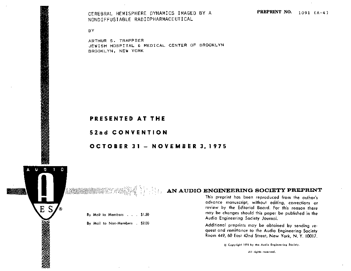
Author (s): Trappier, Arthur S.
Affiliation:
JEWISH HOSPITAL & MEDICAL CENTER OF BROOKLYN, BROOKLYN, NY
(See document for exact affiliation information.)
AES Convention: 52
Paper Number:1091
Publication Date:
1975-10-06
Import into BibTeX
Permalink: https://aes2.org/publications/elibrary-page/?id=2341
(263KB)
Click to purchase paper as a non-member or login as an AES member. If your company or school subscribes to the E-Library then switch to the institutional version. If you are not an AES member Join the AES. If you need to check your member status, login to the Member Portal.

Trappier, Arthur S.; 1975; Cerebral Hemisphere Dynamics Imaged by a Nondiffusable Radiopharmaceutical [PDF]; JEWISH HOSPITAL & MEDICAL CENTER OF BROOKLYN, BROOKLYN, NY; Paper 1091; Available from: https://aes2.org/publications/elibrary-page/?id=2341
Trappier, Arthur S.; Cerebral Hemisphere Dynamics Imaged by a Nondiffusable Radiopharmaceutical [PDF]; JEWISH HOSPITAL & MEDICAL CENTER OF BROOKLYN, BROOKLYN, NY; Paper 1091; 1975 Available: https://aes2.org/publications/elibrary-page/?id=2341